Onion Cell Diagram
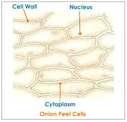
Onion peel cell diagram with label Magnified 40x times 100x microscopy Red onion cell in distilled water 400x red onion cell in salt water
Foods | Free Full-Text | Structural Changes Induced by Pulsed Electric
Beautiful world: onion cells Onion cell red osmosis water distilled lab salt 400x vacuole do studylib The science scoop: onion cell lab
Bulb allium cepa bulbs
Onion cell cells diagram structure 2010 biology microscopic occupies september uses introductionOnion cells microscope hi-res stock photography and images Onion cellsOnion cells under a microscope.
Label cells procedureNcert class 9 science lab manual Onion epidermal epidermis chromosomes chromosome paintingvalleyStaining of onion cell nuclei – microbehunter microscopy.

Biology help online: september 2010
Cells cheek ncert microscope blotting cbsetuts cbseOnion cell diagram drawing Onion cells beautiful worldOnion cells microscope blue methylene stained under observation umberto flickr.
Cebola zwiebel microscope cipolla photomicrograph micrografia micrografo mikrograph resolution microscopio pino legnoOnion cell diagram drawing Onion cells under microscopeOnion skin under microscope 400x.

Onion cell cells microscope micrograph microscopic alamy stock skin magnification section high allium cepa mitosis root
Onion cell cells lab staining nuclei microscope nucleus under stained skin dna simple look power experience class there students tissueOnion cell 400x lab microscope under labeled cells structure scoop science looked Onion microscope epidermis membrane onions biology.
.


Onion Peel Cell Diagram With Label - itsessiii

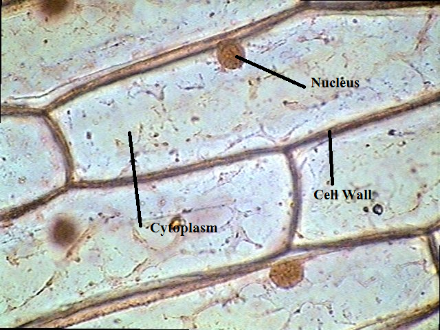
Onion Cells under Microscope

Red onion cell in distilled water 400X Red onion cell in salt water

The Science Scoop: Onion Cell Lab

Onion Cell Diagram Drawing - lana1970

Beautiful World: Onion cells

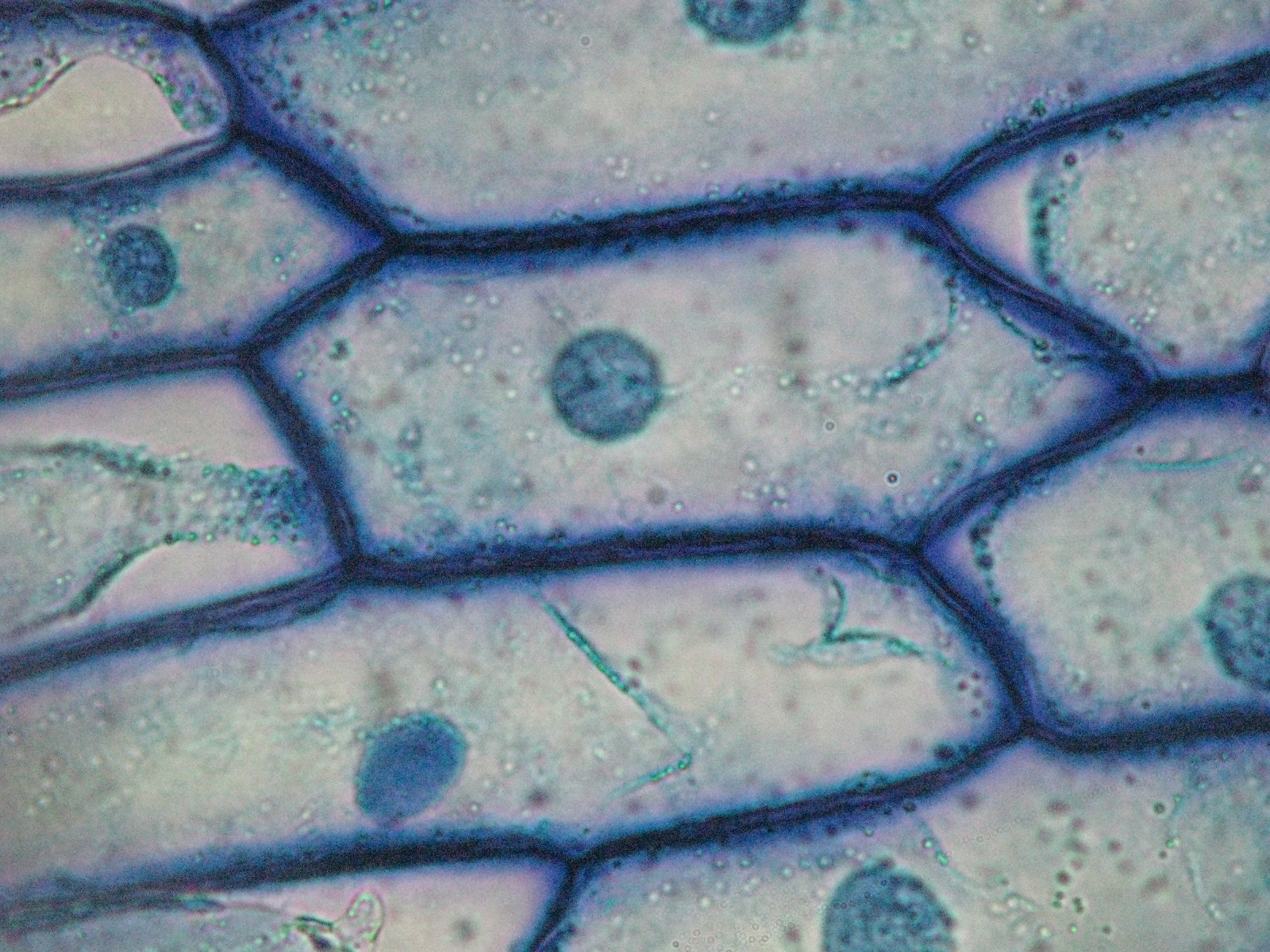
Staining of Onion Cell Nuclei – Microbehunter Microscopy

NCERT Class 9 Science Lab Manual - Slide of Onion Peel and Cheek Cells

Onion Skin Under Microscope 400x | Things Under a Microscope